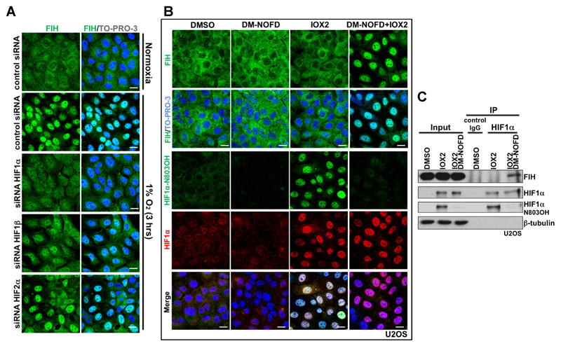
Figure 2. Nuclear entry of FIH is mainly HIF1α-dependent, and requires inhibition of FIH enzymatic activity.
(A) Immunofluorescence staining of FIH (green) in U2OS cells with indicated treatments. U2OS cells were transfected with the indicated siRNA for 3 days, followed by culture in normoxia (20 % O2) or 3 hours’ hypoxia (1 % O2). TO-PRO-3 (blue) was used to stain nuclei. Scale bar: 20 µm.
(B) Immunofluorescence staining of FIH (green), HIF1α N803OH (green) and HIF1α (red) in U2OS cells treated with DMSO, DM-NOFD (1 mM), IOX2 (0.25 mM), or DM-NOFD (1 mM) plus IOX2 (0.25 mM) for 3 hours. TO-PRO-3 (blue) was used to stain nuclei. Scale bar: 20 µm.
(C) Protein levels of FIH, HIF1α and HIF1α N803OH in U2OS cells with indicated treatments. β-tubulin was used as a loading control. Total cell lysates from U2OS cells with indicated treatments were immunoprecipitated with an anti-HIF1α antibody.

