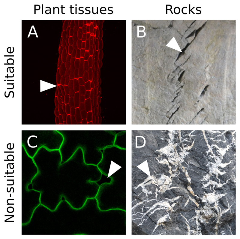
Figure 1. Suitable image type.
A. Z-projection (maximal intensity) of a confocal image stack from a propidium iodide stained light-grown hypocotyl from the qua1-1 mutant with cell adhesion defects (see Verger et al., 2018). B. Cracks in rocks (credit photo: Pierre Thomas). In A and B the cracks are marked by a high pixel intensity contrast and form closed domains. C. Z-projection (maximal intensity) of a confocal image stack from a qua1-1 pPDF1::mCit:KA1 (plasma membrane reporter) cotyledon epidermis (see Verger et al., 2018). Although such fluorescent reporter can provide suitable images for our analysis, in this particular case the contrast between the cracks and the cell content is too low to allow a segmentation of the cracks with our pipeline. D. Picture of cracks in rocks (credit photo: Pierre Thomas). In this case the cracks do not form closed domains as most of them overlap. In addition, the pixel intensity is very variable throughout the picture such that some cracks do not exhibit a strong differential in pixel intensity. White arrowheads point to examples of cracks or cell separations in these images.

