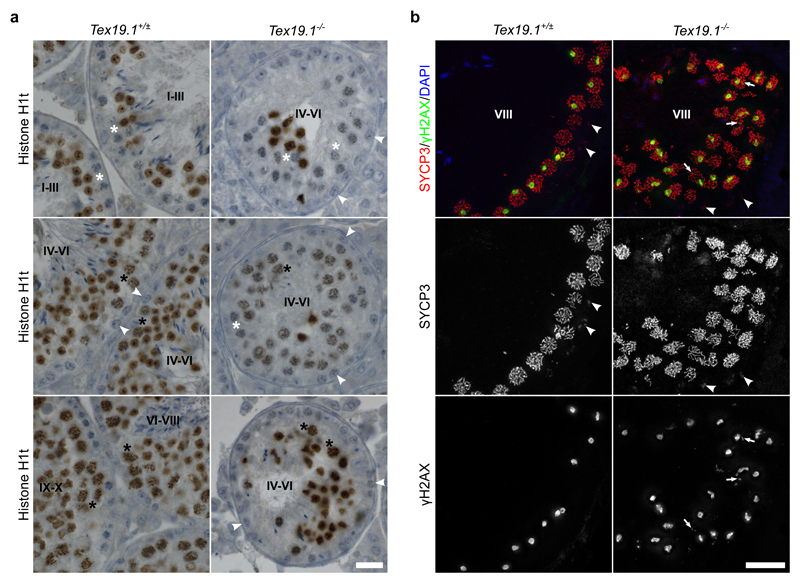
Fig. 5. Features of Early Pachytene Persist in Late Stage Tex19.1-/- Seminiferous Tubules.
a Immunohistochemistry for histone H1t (brown) in Tex19.1+/± and Tex19.1-/- testis sections counterstained with hematoxylin. Testis tubules were staged on the basis of the types of spermatocyte and spermatogonia present (Ahmed and de Rooij 2009). Tubule stages are indicated with Roman numerals and example histone H1t-negative pachytene spermatocytes (white asterisks), histone H1t-positive pachytene spermatocytes (black asterisks), and B spermatogonia (white arrowheads) are annotated. Tubules containing B-spermatogonia (late stage IV to early stage VI) only contained H1t-positive pachytene spermatocytes in Tex19.1+/± testis sections, but two thirds of tubules containing B spermatogonia from Tex19.1-/- testis had some H1t-negative pachytene spermatocytes. 10 tubules containing B spermatogonia were scored for each of three mice for each genotype. Tex19.1-/- tubules have fewer post-meiotic spermatids and smaller diameters than control Tex19.1+/± tubules (Öllinger et al. 2008). Scale bar 20 μm. b Immunofluorescence for the synaptonemal complex component SYCP3 (red) and the DNA damage marker γH2AX (green) in Tex19.1+/± and Tex19.1-/- testis sections counterstained with DAPI (blue) to visualise DNA. 2D projections of deconvolved z-stacks are shown. Grayscale images of SYCP3 and γH2AX are shown below the merged image. Preleptotene spermatocytes (arrowheads) in stage VIII tubules were identified by aggregates of SYCP3 and a lack of γH2AX in spermatocytes around the edge of the tubules (Mahadevaiah et al. 2001; Nakata et al. 2015). γH2AX staining was assessed in the strongly SYCP3-positive pachytene spermatocytes lumenal to the preleptotene spermatocytes. γH2AX was typically present in a large domain likely corresponding to the sex body in these cells, and, in approximately 30% of stage VIII tubules from Tex19.1-/- testes, was also present as additional nuclear foci (arrowheads) in the pachytene spermatocytes. Tex19.1-/- tubules have fewer post-meiotic spermatids and smaller diameters than control Tex19.1+/± tubules (Öllinger et al. 2008). Scale bar 20 μm.

