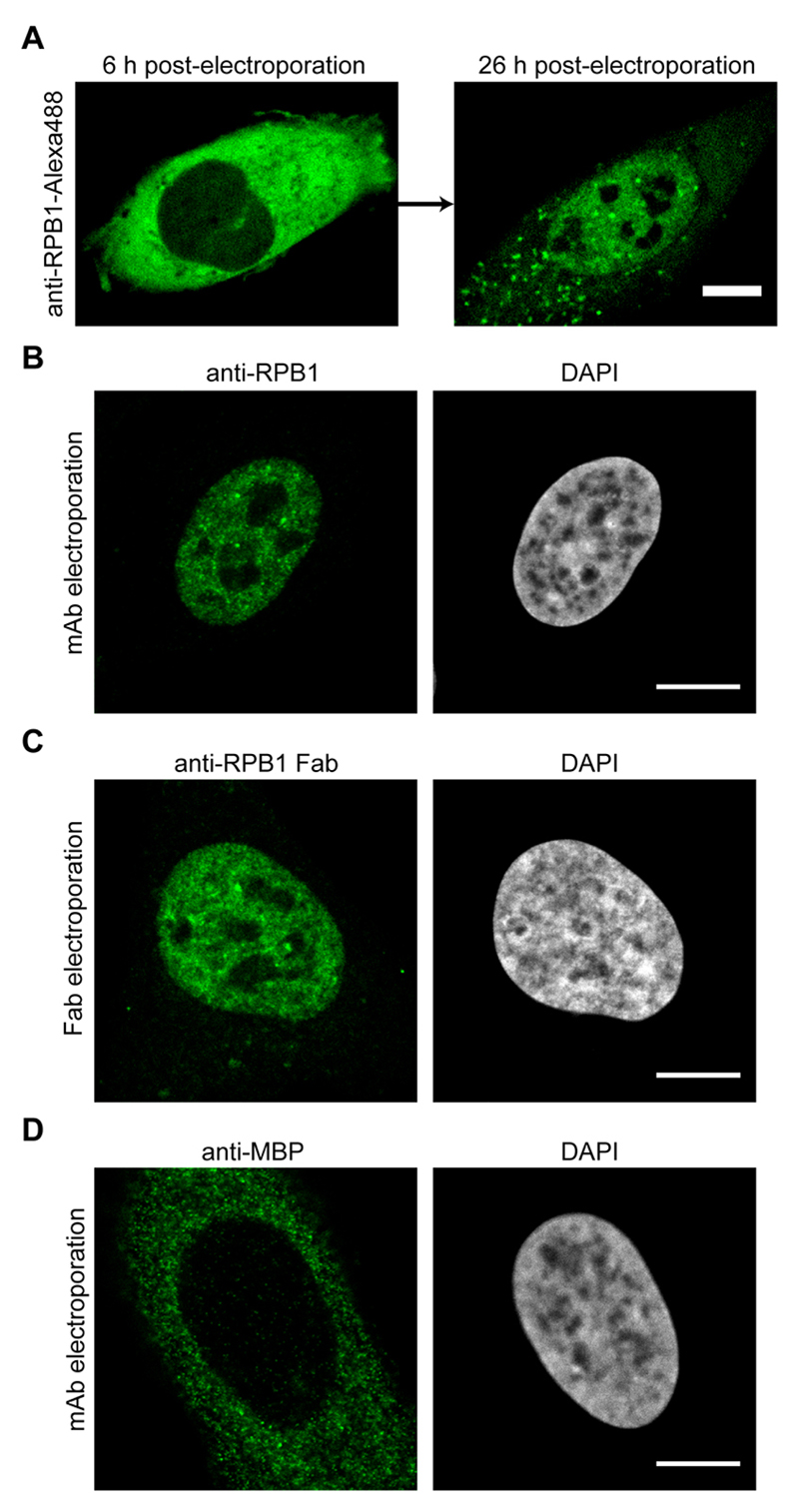
Figure 3. Imaging results after the electroporation of antibodies or Fabs.
A. U2OS cells were electroporated with anti-RPB1-Alexa488 labeled antibodies and the same cell was imaged by confocal microscopy 6 h and 26 h post-electroporation. The transport of the labeled antibody from the cytoplasm into the nucleus can be detected. B. Same as in (A), but the cells were fixed 24 h after electroporation and the antibody labeled RPB1 was visualized using confocal microscopy. C. Electroporation as in (A), but this time anti-RPB1-Alexa488 Fab fragments were transduced. The cells were fixed and imaged 6 h post-electroporation by confocal microscopy and specific nuclear staining for RPB1 can be observed. D. U2OS cells were electroporated with an antibody against MBP (maltose binding protein), which is not present in human cells. The cells were fixed 24 h after electroporation and the localization of the antibody was visualized using confocal microscopy. Even after 24 h, the antibody stays in the cytoplasm as it has no target protein for the transport into the nucleus. Scale bars = 10 μm.

