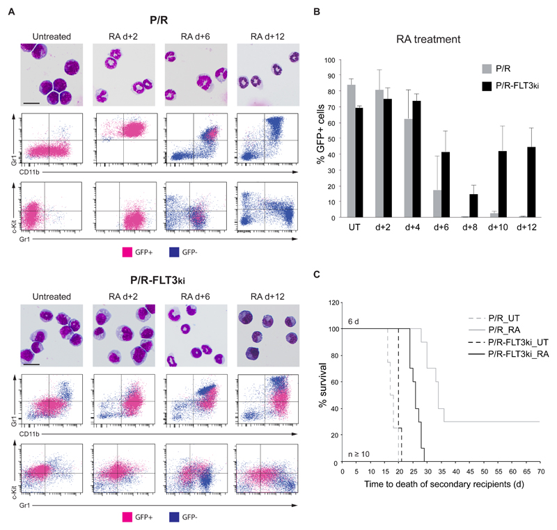
Figure 2. Cell differentiation and blast clearance in P/R and P/R-FLT3ki APL upon ATRA treatment.
(A) ATRA-induced differentiation of P/R (top) and P/R-FLT3ki (bottom) bone marrow cells as assessed by May-Grünwald-Giemsa (MGG) staining (scale bar 10 μm). FACS analysis after 2-, 6- and 12-d of in vivo ATRA treatment. GFP-positive leukemic cells (pink) and GFP-negative normal cells (blue) are shown. (B) Percentages of GFP-positive APL cells in bone marrow of untreated (UT) and ATRA-treated P/R (grey bars) and P/R-FLT3ki (black bars) mice at 2- to 12-d post-treatment. Data are expressed as mean ± s.d. of two independent experiments. (C) Survival of secondary recipients transplanted with total APL bone marrow cells from ATRA 6-d-treated primary APL mice in both models (n ≥ 10 for each model).

