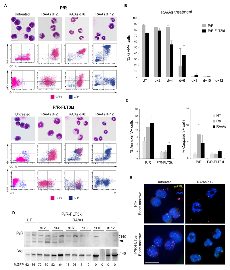
Figure 5. Arsenic drives elimination of FLT3ki APLs.
(A) ATRA/As-induced differentiation of P/R (top) and P/R-FLT3ki (bottom) bone marrow cells as assessed by MGG staining (scale bar 10 μm) and FACS analysis after 2-, 6-, 8- and 12-d of in vivo treatment. GFP-positive leukemic cells (pink) and GFP-negative normal cells (blue) are shown. (B) Percentages of GFP-positive APL cells in bone marrow of untreated (UT) and ATRA/As-treated P/R (grey bars) and P/R-FLT3ki (black bars) mice at 2- to 12-d post-treatment. (C) Proportion of Annexin-V (left) and Active Caspase-3 (right)-positive spleen cells in APL mice treated for 4 days as indicated. (D) Western blot of PML/RARA (P/R, arrowheaded) and vinculin (Vcl) expression in the bone marrow of P/R-FLT3ki APL untreated (UT) and ATRA/As-treated mice from 2- to 12-d. (E) PML NB reformation assessed by immunofluorescence analysis of murine PML (green) and human PML/RARA (red) with DAPI (blue) in bone marrow cells of P/R and P/R-FLT3ki APL mice treated with ATRA/As after 2-d (scale bar 10 μm).

