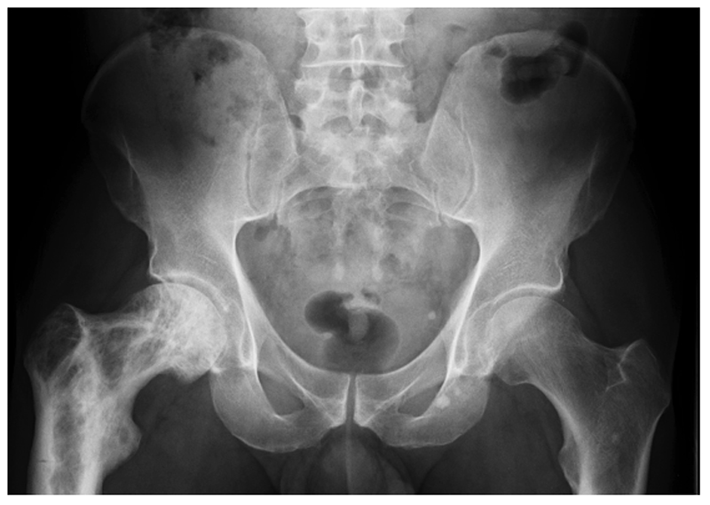
Figure 1. X-ray features of PDB.
Pelvic radiograph from a patient with PDB affecting the upper right femur showing alternating areas of osteolysis and osteosclerosis in the greater and lesser trochanters and femoral neck; loss of distinction between the cortex and medulla in the upper femur; bone expansion and deformity of the affected femur; and a pseudofracture on the lateral aspect of the femur opposite the lesser trochanter.

