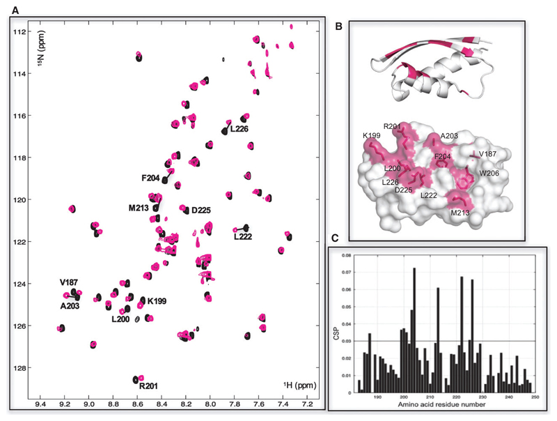
Fig. 2.
The MYC:MAX bHLHZip dimer binds to a conserved pocket on INI1/hSNF5 RPT1. (A) 1H,15N HSQC spectra of RPT1 without (black) and with (magenta) the addition of unlabeled MYC:MAX bHLHZip dimer (ratio 1 : 2). Residues undergoing chemical shift changes more than the standard deviation (SD) are labeled in black. (B) Cartoon (top) and molecular surface representation (bottom) of RPT1 showing the residues that undergo chemical shift changes more than the standard deviation are highlighted in magenta and labeled. The chemical shift changes of W206 are for the aromatic proton of the side chain (changes in chemical shifts of the backbone are below the threshold). (C) Diagram showing the differences in chemical shifts induced by binding of the MYC:MAX dimer to the 15N-labeled INI1 RPT1. The black line indicates the calculated standard deviation.

