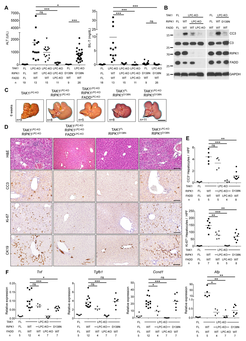
Fig. 3. RIPK1 kinase activity drives cholestasis but is largely dispensable for hepatocellular damage in TAK1LPC-KO mice.
(A) Serum levels of ALT and total Bilirubin in 6-week-old mice with the indicated genotypes. (B) Immunoblot analysis of whole liver lysates from 6-week-old mice with the indicated genotypes. GAPDH was used as loading control. (C) Representative liver images from 6-week-old animals with the indicated genotypes. (D) Representative images of liver sections from 6-week-old mice with the indicated genotypes stained with H&E or immunostained for CC3, Ki-67 and CK19. (E) Quantification of CC3 and Ki-67 immunostaining shown in D. (F) qRT-PCR gene expression analysis in liver samples of mice with the indicated genotypes. Graphs show relative mRNA expression normalized to Tbp. The number of mice analyzed (n) is indicated in every graph. All graphs show mean values of the individual data points. *p <0.05, **p <0.01, ***p <0.005. Bars: (C) 1 cm; (D) 100 μm.

