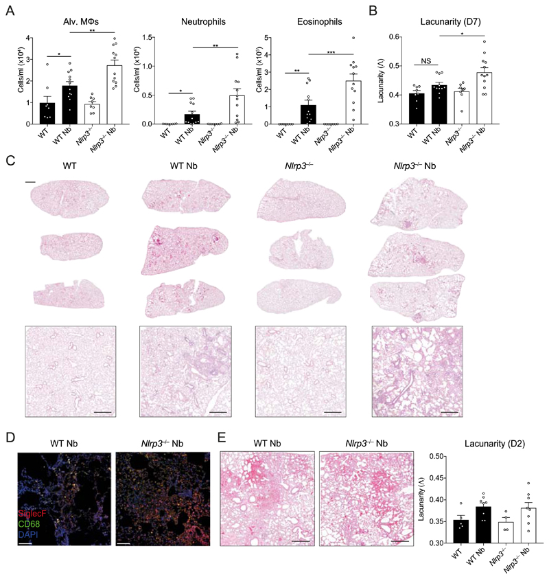
Figure 3. NLRP3 regulates lung tissue repair following N. brasiliensis infection.
WT and Nlrp3−/− mice were infected with N. brasiliensis (Nb) and (A) day 7 post-infected BAL alveolar (alv.) Mφ (CD11b−Siglec-F+CD11c+), neutrophil (CD11b+Siglec-F−Ly6G+) and eosinophil (CD11b+Siglec-F+) absolute numbers were measured by flow cytometry. Haematoxylin/eosin staining was performed on lung sections and imaged followed by (B) quantification of lacunarity (Λ) to assess lung damage on day 7. (C) Representative image insets are shown on top with low magnification scans of entire left lung lobes from individual mice shown below (top panel scale bar = 1000 µm, bottom panel scale bar = 500 µm). (D) Immunofluorescence staining of lung inflammatory foci with SiglecF (red), CD68 (green), and DAPI (blue) (scale bar = 100 µm). (E) Representative day 2 post-infection lung sections and lacunarity. Data were pooled (A–B, E; mean ± s.e.m.) from 3 individual experiments with 3-5 mice per group (per experiment). *P<0.05, **P<0.01, ***P<0.001 (one-way ANOVA and Tukey-Kramer post hoc test).

