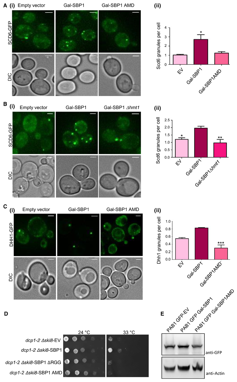
Fig. 4. Sbp1 AMD mutant fails to perform the role of Sbp1 in decapping.
(A)i) Galactose-inducible wild type or AMD mutant of Sbp1 was transformed into Scd6-GFP strain. Cells were grown in SD-URA with 2% glucose up to 0.3–0.35 OD600 followed by growth in 2% galactose containing media for 2 h. (ii) Quantitation of granule count for experiments (n = 4) performed in A using mean SEM. At least 150 cells were counted per experiment. P value = 0.035, calculated using two-tailed paired t-test. (B)(i) Ability of Sbp1 to induce Scd6-GFP granule was compared in WT and Δhmt1 background. (ii) Quantitation of Scd6-GFP granule for experiment (n = 4) performed in (B)(i) using mean SEM, P value = 0.0085, calculated using two-tailed paired t-test. (C)i) Galactose-inducible wild type or AMD mutant of Sbp1 was transformed into Dhh1-GFP strain. Cells were grown in SD-URA with 2% glucose up to 0.4 OD600 followed by growth in 2% galactose containing media for 4 h. (ii) Quantitation of granule count for experiments (n = 3) performed in (C)(i). For quantitation a threshold intensity of 1500 was set up for all the images and granules with intensities more than 1500 are counted and plotted. At least 100 cells were counted per experiment using mean SEM. P-value for difference in Dhh1 granule between WT and AMD mutant is =0.0030, calculated using two-tailed paired t-test. *Denotes statistical significance. (D) Plasmid expressing WT and mutants of Sbp1 were transformed into dcp1-2 Δski8 strain. Growth assay was performed by spotting these cultures on SD-LEU plates and incubating them at 25 °C (permissive temperature) and 33 °C (non-permissive temperature). Image for 24 °C plate was taken on day 2 while 33 °C plates were imaged on day 4. (E) Pab1-GFP levels upon overexpression of galactose-inducible SBP1. Expression of Sbp1 or its mutants in Pab1-GFP strain was induced for 4 h followed by pelleting and breaking open as described in methods section. Pab1 was detected using anti-GFP. Actin was used as loading control.

