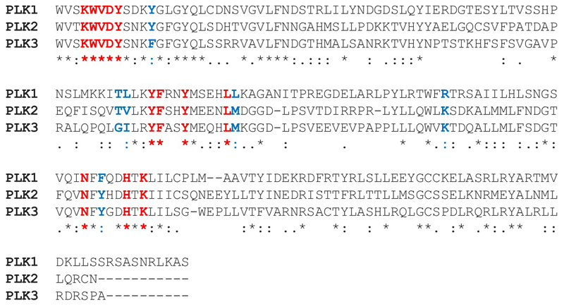
Figure 5.
Sequence alignment of the PBDs of the PLK family. Sequences of human PLK1 residues 410-603, PLK2 residues 503-685, and PLK3 residues 463-646 were taken from UniProt. PLK4 and PLK5 were excluded from the analysis as they have markedly different structures and functions. Alignment was performed by Clustal Omega using default parameters. Residues in the phosphopeptide binding groove and cryptic hydrophobic pocket are highlighted in bold. Residues in the phosphopeptide binding groove and cryptic hydrophobic pocket with complete conservation across the PLK family are in red and those which differ are in blue.

