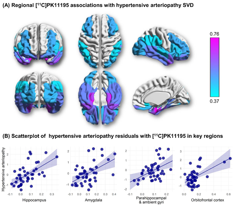
Figure 4. Associations between regional [11C]PK11195 binding and hypertensive arteriopathy (n=42).
(A) β-weight brain mapping of associations between regional [11C]PK11195 binding and hypertensive arteriopathy. Coloured overlay of the Hammers atlas represents statistically significant elevation of [11C]PK11195 binding with hypertensive arteriopathy score controlling for sex, age, education, ACE-R score, and scan interval, and corrected for multiple comparisons. Colour gradient represents the strength of association (standardized β weights), increasing in magnitude from light blue to fuchsia. (B) Scatterplots of relationships between hypertensive arteriopathy (y-axis) and [11C]PK11195 binding in key regions of interest (x-axis). Hypertensive arteriopathy values are residuals adjusting for sex, age, education, ACE-R, and scan interval.

