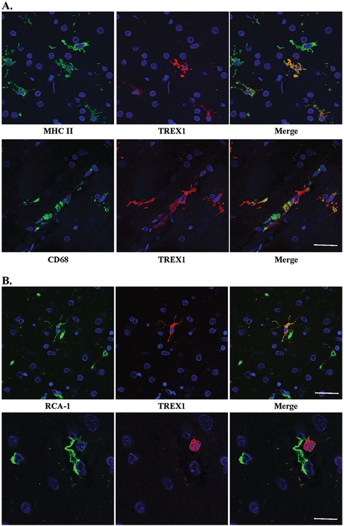
Figure 4.
TREX1 positive cells are microglia and/or macrophages. A. Dual staining of frozen tissue from a case of RVCL with anti-MHC II (top panel, green) and anti-CD68 (bottom panel, green), microglial markers, and anti-TREX1 (red). Nuclei are counterstained with TO-PRO-3 (blue). Scale bar represents 28 μm. B. Two examples (upper and lower panels) of dual staining of formalin-fixed, paraffin-embedded human brain tissue from a normal control with RCA-1 (left panel, green), a microglial/macrophage and endothelial cell marker, and anti-TREX1 (red). Nuclei are counterstained with TO-PRO-3 (blue). Some cells stained with RCA-1 but not TREX1 are endothelial cells based on their morphology. Scale bars represent 28 μm on top panel and 14 μm on bottom panel.

