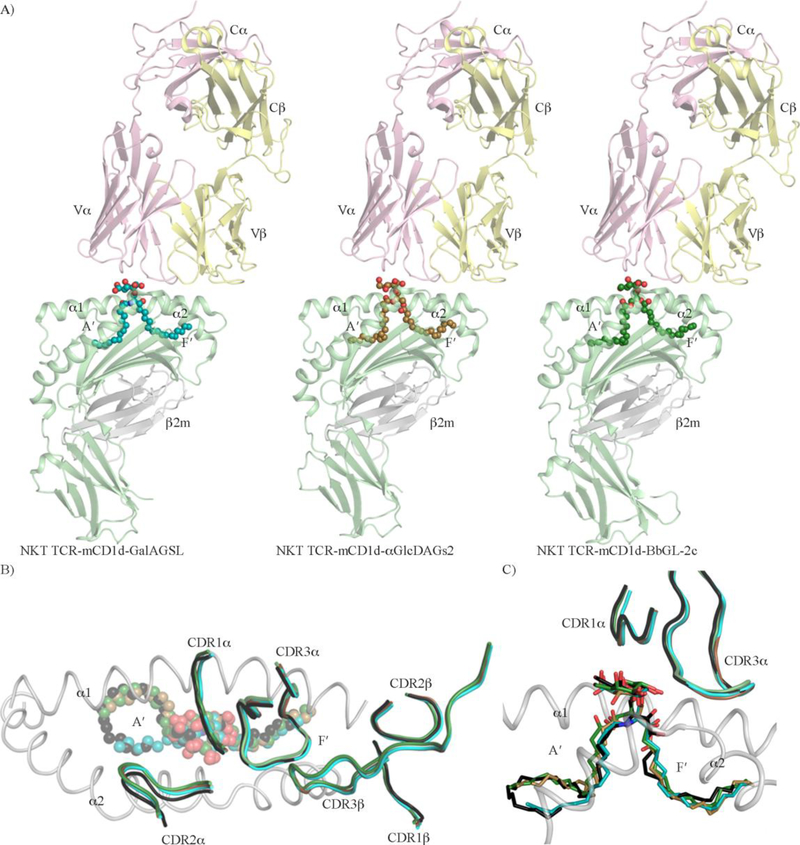
Figure 4. Molecular recognition of CD1d presenting microbial lipid-based Ags by NKT TCR.
A) Crystal structures of NKT TCR-mCD1d-GalAGSL (left panel), NKT TCR-mCD1d-αGlcDAGs2 (middle panel), and NKT TCR-mCD1d-BbGL-2c (right panel) ternary complexes. The mCD1d and β2-microglobulin molecules are coloured in light green and light grey, respectively. The NKT TCRα and TCRβ are coloured in pink and yellow, respectively. The microbial glycolipids GalAGSL (cyan), αGlcDAGs2 (brown) and BbGL-2c (dark green) are shown as spheres. B) View from the top of an overlay of the three NKT TCR-mCD1dmicrobial lipids crystal structures and NKT TCR-mCD1d-α-GalCer. For clarity, only the CDR loops are shown and coloured as for the respective lipids in each structure, GalAGSL (cyan), αGlcDAGs2 (brown), BbGL-2c (dark green) and α-Galactosylceramide (α-GalCer) (black). The lipids are shown as spheres. C) Overlay of three NKT TCR-mCD1d-microbial lipid crystal structures and NKT TCR-mCD1d-α-GalCer. For clarity, only the CDR1α and CDR3α loops are shown and coloured as for the respective lipids in each structure, GalAGSL (cyan), αGlcDAGs2 (brown), BbGL-2c (dark green) and α-Galactosylceramide (α-GalCer) (black). The lipids are shown as sticks.

