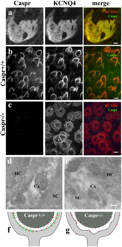
Figure 3. Localization of KCNQ4 at the calyceal membrane depends on Caspr.
(a) Confocal optical section grazing the calyx- type I hair cell (from adult rat ampulla) contact region shows perfectly matched patterns of distribution of the immunofluorecence for Caspr (red) and KCNQ4 (green) indicating that these two proteins are co-localized. (b, c) Caspr immunoreactivity in ampulla of Caspr+/+ and Caspr-/- at P18. (b) Caspr and KCNQ4 colocalize at the calyceal synapses in Caspr+/+ mice. (c) The labeling for KCNQ4 in the membranes of the Caspr-/- calyx is faint and sparsely distributed. (d, e) Immunogold labeling of KCNQ4 confirms its localization at the internal membranes of the nerve calyx in Caspr +/+ and along both internal and external membranes of the calyx in Caspr-/-. Schematic diagram shows the colocalization of Caspr and KCNQ4 in Caspr +/+ (f) and the redistribution of KCNQ4 in Caspr-/- (g). An yet unidentified binding partner for the Caspr complex on the hair cell membrane is represented in gray (f and g). Scale bar, a, 2 μm; b-c, 10 μm; d-e, 200 nm.

