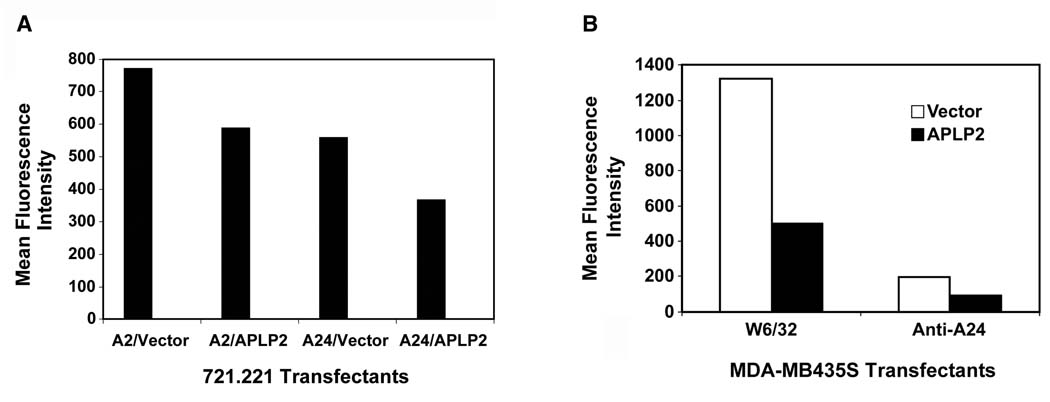
Figure 6.
(A) APLP2 transfectants of 721.221-A24 and 721.221-A2 cells had decreased cell surface HLA class I expression compared to vector-only transfectants. The bar graph displays the cell surface expression of HLA-A24 or HLA-A2 molecules at the surface of 721.221 cells transfected for 24 hours with vector only (pCMVTag4A) or with an APLP2 cDNA in pCMVTag4A. The cells were stained with either anti-A2 antibody (BB7.2) or anti-HLA-A24 antibody, washed and stained with phycoerythrin-labeled secondary antibody, and analyzed on a BD FACSCalibur. Separate results indicating that increased expression of APLP2 reduced HLA-A24 surface expression on transfected 721.221 cells were obtained in a separate experiment. (B) APLP2 transfectants of breast carcinoma MDA-MB435S cells had decreased cell surface HLA class I expression compared to vector-only transfectants. The bar graph displays the cell surface expression of total (W6/32+) HLA class I molecules or HLA-A24 molecules at the surface of MDA-MB435S cells stably transfected with vector only (pCMVTag4A) or with an APLP2 cDNA in pCMVTag4A. The cells were stained with either W6/32 or with anti-HLA-A24 antibody, washed and stained with phycoerythrin-labeled secondary antibody, and analyzed on a BD FACSCalibur. Separate results indicating that elevated expression of APLP2 reduced HLA-A24 surface expression on APLP2-transfected MDA-MB435S cells were also obtained in two additional experiments.

