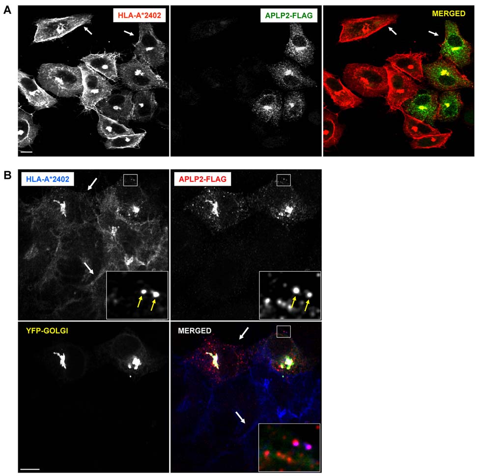
Figure 7.
Elevated expression of APLP2 in HeLa cells reduced the amount of HLA-A24 at the cell surface. (A) HeLa cells stably expressing HLA-A*2402 were transfected with APLP2-FLAG by the use of Effectene. At 24 hours, the cells were fixed by treatment with 4% paraformaldehyde in PBS for 10 minutes, incubated with primary antibodies (mouse anti-HLA-A24 and rabbit anti-FLAG antibodies) in staining solution for 1 hour at room temperature, and washed (3 times in PBS, 5 minutes/wash). The cells were then incubated with fluorochrome-conjugated secondary antibodies (Alexa Fluor 568 goat anti-mouse antibody and Alexa Fluor 488 goat anti-rabbit antibody) in staining solution for 30 minutes at room temperature, and washed again 3 times with PBS for 5 minutes/wash. The arrows at the upper left of the first and third panels point to an area of bright staining of HLA-A*2402 molecules at the surface of HeLa-A24 cells, and the arrows at the upper right of the same panels point to an area of relatively weak staining on a HeLa-A24 cell transfected with APLP2-FLAG. (B) HeLa cells stably expressing HLA-A*2402 were transfected with APLP2-FLAG and YFP-Golgi by the use of Effectene. At 24 hours, the cells were fixed by treatment with 4% paraformaldehyde in PBS for 10 minutes, incubated with primary antibodies (mouse anti-HLA-A24 and rabbit anti-FLAG antibodies) in staining solution for 1 hour at room temperature, and washed (3 times in PBS, 5 minutes/wash). The cells were then incubated with fluorochrome-conjugated secondary antibodies (Alexa Fluor 405 goat anti-mouse antibody and Alexa Fluor 568 goat anti-rabbit antibody) in staining solution for 30 minutes at room temperature, and washed again 3 times with PBS for 5 minutes/wash. Arrows in the larger boxes indicate less HLA-A*2402 at the plasma membrane of a cell transfected with APLP2-FLAG (top arrow) compared to more HLA-A*2402 at the surface of a cell that was not transiently transfected with APLP2-FLAG. Insets depict more highly magnified images of the areas shown in the larger boxes. The arrows within the insets indicate peripheral vesicles in which HLA-A*2402 (blue) and APLP2-FLAG (red) are co-localized (magenta). Blue = HLA class I; red = APLP2-FLAG; yellow = YFP-Golgi; white = merged. For both (A) and (B) images were analyzed on a Zeiss LSM 5 Pascal confocal microscope and bar = 10 µm.

