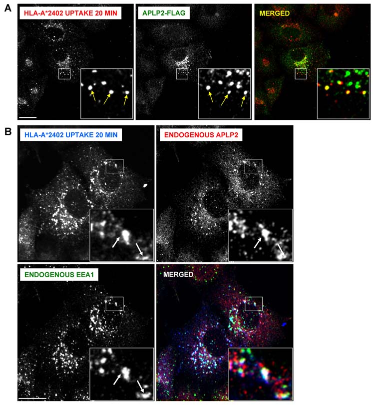
Figure 8.
Endocytosed HLA-A*2402 molecules co-localized with APLP2 in early endosomes. (A) HeLa cells stably expressing HLA-A*2402 were transfected with APLP2-FLAG by the use of the Effectene transfection reagent. At 24 hours, the cells were pulsed with anti-HLA-A24 antibody for 20 minutes. At the end of the pulse period, remaining cell surface-bound antibodies were removed by incubating the cells with stripping solution (0.5% acetic acid, 500 mM NaCl) for 90 seconds, and the cells were fixed by incubation with 4% paraformaldehyde in PBS for 10 minutes. The cells were then incubated in staining solution with rabbit anti-APLP2 serum for 1 hour at room temperature, washed 3 times (5 minutes/wash) with PBS, incubated for 30 minutes at room temperature with Alexa Fluor 568 goat anti-mouse and Alexa Fluor 488 goat anti-rabbit antibodies in staining solution, and washed 3 times with PBS (5 minutes/wash). Red = HLA-A*2402; green = APLP2-FLAG; yellow = merged. Insets represent more highly magnified images of the areas that are in the larger boxes. Arrows within the insets indicate some of the vesicles in which HLA-A*2402 and APLP2-FLAG are co-localized. (B) HeLa cells stably expressing HLA-A*2402 were pulsed with anti-HLA-A24 antibody for 20 minutes. After the pulse period, remaining surface-bound antibodies were removed by treating the cells with stripping solution (0.5% acetic acid, 500 mM NaCl) for 90 seconds, and the cells were fixed by incubation in 4% paraformaldehyde in PBS for 10 minutes. The cells were then incubated in staining solution with Alexa Fluor goat anti-mouse 405 antibody for 30 min. After 3 washes of 5 minutes each with PBS, the cells were stained with rabbit anti-APLP2 serum and anti-EEA1 (IgG1) antibody for 1 hour at room temperature, washed 3 times (5 minutes/wash) with PBS, incubated for 30 minutes at room temperature with Alexa Fluor 568 goat anti-rabbit and Alexa Fluor 488 goat anti-mouse IgG1 antibodies in staining solution, and washed 3 times with PBS (5 minutes/wash). Blue = HLA-A*2402; red = APLP2; green = EEA1; white = merged. Insets represent more highly magnified images of the areas that are in the larger boxes. Arrows within the insets indicate some of the vesicles in which HLA-A*2402, APLP2, and EEA1 are co-localized. For both (A) and (B), the images were analyzed on a Zeiss LSM 5 Pascal confocal microscope and bar = 10 µm.

