
Figure 1. Structures of FhaC and the TPS domain of FhaB.
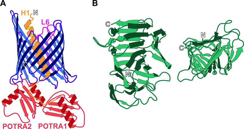
(A) Helix 1 (H1, orange) and loop 6 (L6, fuchsia) are located within the pore of the 16-stranded β barrel (blue) of FhaC when the transporter is in the “closed” state. The POTRA domains (POTRA1 and POTRA2, red) remain periplasmic for selective recognition of the FhaB TPS domain. (B) The TPS domain of FhaB adopts a triangular β-helical structure, shown from the side of the helix (left) and top-down in a C-terminus to N-terminus direction (right). Termini are indicated by outlined letter.