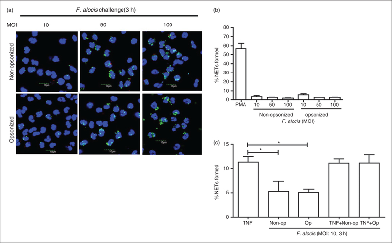
Figure 2.

F. alocis challenge at increasing MOIs fails to induce NET formation by human neutrophils. Neutrophils were challenged with CFSE-labeled viable non-opsonized F. alocis (MOI 10, 50, and 100) or with opsonized F. alocis (MOI 10, 50, and 100) for 180 min. Following infection, cells were fixed and immunostained using Abs directed against MPO (AlexaFluor647), DNA was stained with DAPI, and imaged for NET immunofluorescence by confocal microscopy. (a) Representative confocal images (from 3 independent experiments of 100 quantified cells per experiment) of neutrophils challenged with CFSE-labeled non-opsonized F. alocis (MOI 10–50–100) for 180 min or neutrophils challenged with CFSE-labeled opsonized F. alocis (MOI 10–50-100) for 180 min. CFSE-F. alocis (shown in green); neutrophil nucleus/DNA-DAPI (shown in blue); neutrophil MPO (AlexaFluor647 shown in red), merge image: NET formation. (b) Quantification of percentage of NETs formed using ImageJ analysis. Data are expressed as means of % NETs formed +/− SEM from 3 independent experiments. (c) Neutrophils were stimulated with TNF-α (10 min) or challenged with CFSE-labeled non-opsonized F. alocis (MOI 10), or CFSE-labeled opsonized F. alocis (MOI 10) for 180 min, or pretreated with TNF-α (10 min) followed by CFSE-labeled non-opsonized F. alocis (MOI 10) challenge, or CFSE-labeled opsonized F. alocis (MOI 10) challenge for 180 min. Following infection, cells were fixed and immunostained using Abs directed against MPO (AlexaFluor647), DNA stained with DAPI, and imaged for NET immunofluorescence by confocal microscopy. Quantification, using ImageJ analysis, of percentage of NETs formed from neutrophils at the different experimental conditions detailed above was performed. Data are means ± SEM from four independent experiments. *P < 0.05
