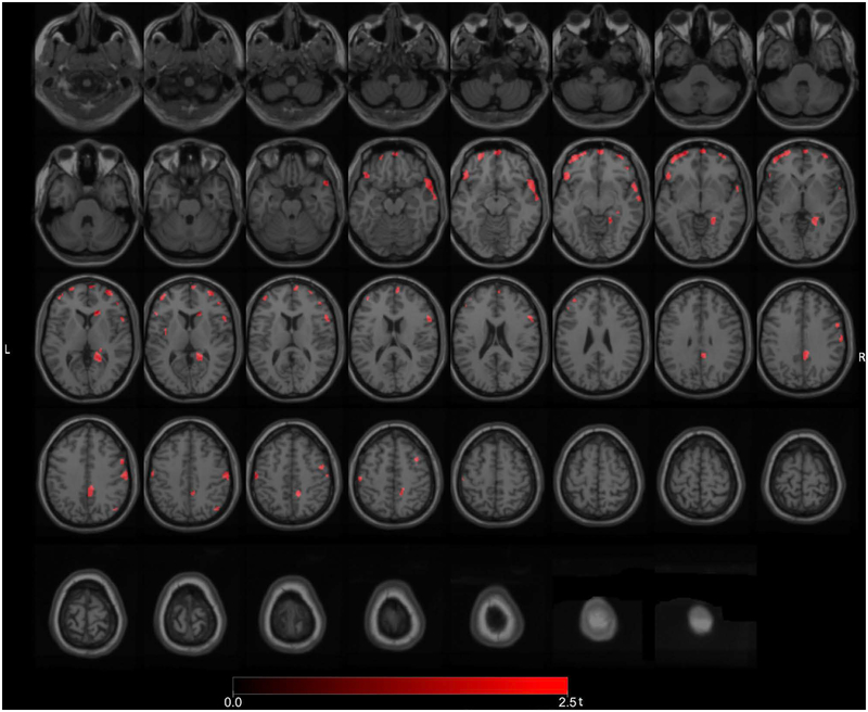
Figure 3.
Post hoc exploratory voxel-based SPM analysis comparing the subset of PD fallers without FoG to PD non-fallers also without FoG. There were more isolated reductions in the right LGN, right caudate nucleus, right premotor cortex, right frontal eye field, right temporopolar cortex, right lateral temporal, right posterior cingulum, right proximal lingual gyrus and bilateral prefrontal regions. Additional reductions were seen in the right more than left sensorimotor cortices.

