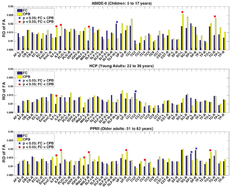
Figure 7:

Reproducibility of diffusion FA measure of anatomical fiber tracts identified from the test-retest dMRI data using the fiber clustering (FC) and cortical-parcellation-based (CPB) methods. A low relative difference (RD) value represents a high test-retest reproducibility. The tracts with significantly lower mean relative difference scores using the fiber clustering method are annotated with a red asterisk, while the tracts with significantly lower mean relative difference scores using the cortical-parcellation-based method are annotated with a blue X.
