
FIG. 12.
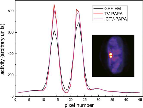
One-channel-wide line profiles through reconstructed transaxial images of clinical Tc-99 m Sestamibi parathyroid scan image shown in Fig. 10. The location of the profile is shown in the inset. Penalty weights were set as: the TV-PAPA method: λ = 2, the ICTV-PAPA method: λ1 = 2, λ2 = 2.