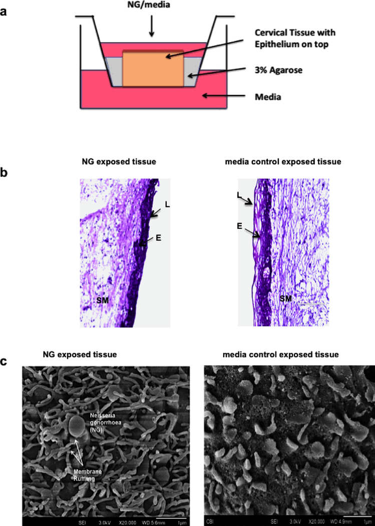
Figure 1: Ecto-cervical tissue based organ culture model to study physical responses and induction of inflammatory cytokines from the cervical tissues in presence of NG.
(a) Transwell with ecto-cervix tissue surrounded with agarose, NG was added to the apical surface of the tissue, with media at the top and bottom well, and incubated for 24 or 48 hours. (b) H&E staining on ecto-cervical tissues exposed to NG or control media in the organ culture for 24 hours. Images were obtained by bright field microscopy (E: Epithelium; L: Lumen of ecto-cervix; SM: Sub-mucosa of the ecto-cervix). Magnification for viewing these ecto-cervical tissue sections was 20X. Each donor had 2–3 control and 2–3 NG exposed biopsies. with 5–10 random images obtained from each biopsy. (c) Membrane ruffling in presence of NG characterized by microvilli projection observed in cervical biopsy post 24 hours exposure to NG under SEM, with a magnification of 20,000X.

