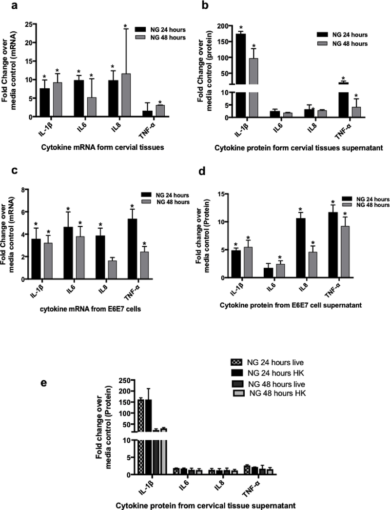
Figure 2: Cellular responses induced by NG in cervical tissues and cervical tissue derived cell lines.
Cervical tissue biopsies exposed to NG showed elevation in cytokine levels. Inflammatory cytokines (a)mRNA and (b) protein at 24 hours and 48 hours compared to media control showed high fold changes in IL-1β (5–10 fold in mRNA and 100–200 fold in protein) and TNF-α (2–5 fold in mRNA and 5–10 fold in protein). There was a significant increase in the level of IL-1β and TNF-α protein in supernatant at both time points though we did not observe a significant increase in TNF-α mRNA at 24 hours. The (c) mRNA profile and (d) secreted cytokine protein profile of the E6/E7 Cells upon exposure to NG showed a similar increase in cytokine responses as in the tissues with an significant increase of 3 and 5 folds (IL-1β) and 5 and 10 fold (TNF-α )of cytokines at the mRNA(Fig. 2c) and protein levels respectively. Ecto-Cervical tissue biopsies exposed to either (e) live NG or heat killed NG showed no difference in cytokine response. Bars represent mean ± standard dev of three independent experiments with different donors. Each donor had 2–3 control and 2–3 NG exposed biopsies for each experimental set. Experiemnts with E6/E7 cells were carried out in triplicates. P=<0.05 was considered to be statistically significant compared to the control tissues for these fold changes analyzed by one sample students T test.

