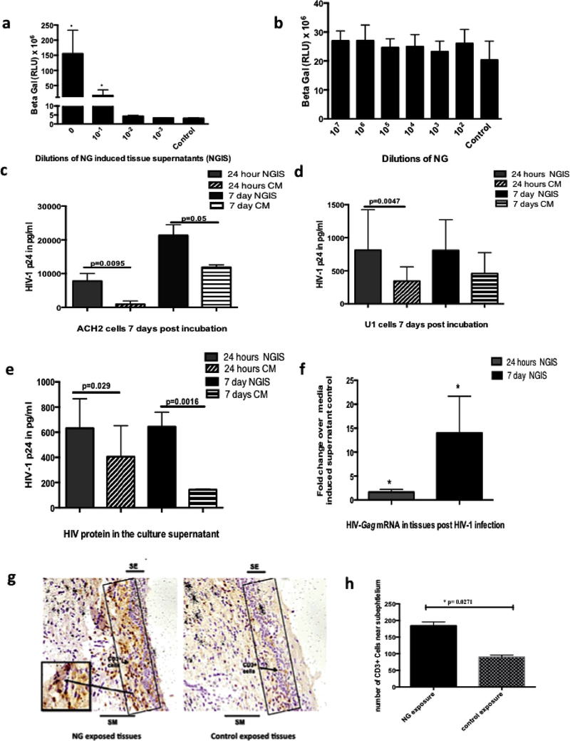
Figure 3: Evaluation of the role of NG and NGIS on the replication and transmission of HIV-1.
(a)Dose dependent increase in transcription of HIV-1LTR in the TZM-bl demonstrated by increased beta-galactosidase activity by NGIS compared to CM. (b) NG per se did not show any stimulation of the HIV-LTR activity over control media (CM). NGIS induced higher replication and production of virus particles from latently infected cell lines (c) ACH2 cells and (d) U1 cells compared to CM at both 24 hours and 7 days. All cell studies were carried out in three independent experiments with 3 replicates in each and were analysed using parametric t tests. (e) NGIS increased the transmission of HIV-1 (HIVBAL 300uL of TCID50 of 10 6)across the mucosa as demonstrated by the transmitted virus (HIV p24) and (f) increase in HIV-Gag mRNA in the tissues. The p-value was calculated as significant using unpaired t test of equal variance because of unequal number of bipsy per condition. (g) Increased localization of CD3+ cells observed in the sub-epithelium in NG exposed tissues over CM exposed tissues. Figure is a representative image at 200X magnification. (h) Quantitation of immuno-stained CD3+ cells showed a statistically significant increase in CD3+ T cells on NG exposed tissue compared to CM exposed tissues. p=<0.05 was considered as significant in all cases. For the CD3+ tissue stain, experiments were carried out in 2–3 biopsies from tissues of 3 donors and non-parametric paired Wilcoxon signed ranked test was used in the analysis of this data.

