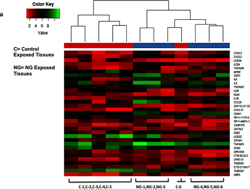
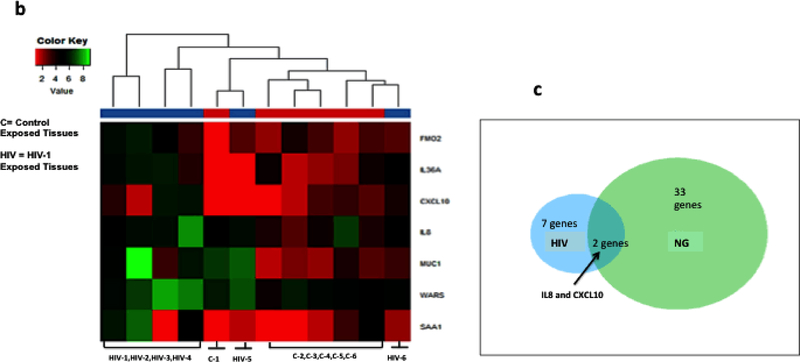
Figure 4: Transcriptome analysis of cervical epithelium exposed to NG and HIV-1 identified up-regulated cellular genes.
Heat Map showing transcriptome analysis of epithelial layer of the cervix using next- generation sequencing of tissue epithelium exposed to (a)NG (n=6) and (b) HIV-1 (n=6). NG depicts Neisseria numbered 1–6, HIV-1 is also numbered 1–6. C depicts tissues exposed to control supernatant and numbered as 1–6. Figure shows only the 6 tissues which were subjected to transcriptome analysis and genes which were found to be upregulated. Ven-diagram showing (c) common genes CXCL10 and IL8 expressed by NG and HIV-1 exposure on the cervical epithelium. The expression of the genes in (a) NG exposed and (b) HIV-1 exposed ectocervical epithelia was at least 2-fold difference with false discovery rate (FDR) <0.05 compared to the controls.


