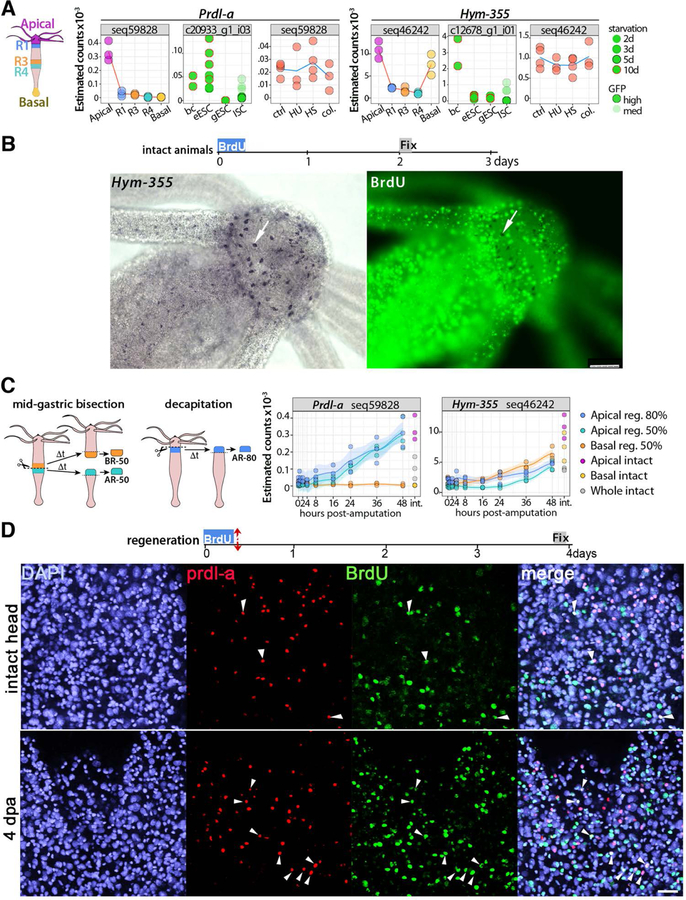
Fig.2. Homeostatic and regeneration-induced de novo neurogenesis in slow aging Hydra vulgaris.
(A) Expression profiles of the neurogenic genes Prdl-a and Hym-355 as detected in animals of the heat-sensitive Hv_sf-1 strain through three different RNA-seq approaches performed (a) on tissues from slices of the body column when distinct regions (Head, R1, R3, R4, Foot) were dissected before RNA extraction (see the scheme on the left that indicates the five regions); (b) on distinct cell types, i.e. ectodermal epithelial stem cells (eESC), gastrodermal epithelial stem cells (gESC) and interstitial stem cells (ISCs) isolated by flow-cytometry; (c) on the central body column of animals exposed to drugs (HU, colchicine) or Heat-shock (HS). All transcriptomic procedures are described in (Wenger et al., 2016). (B) Lack of BrdU+ neurons expressing Hym-355 in animals exposed to BrdU for four hours and fixed four days later. Scale bar: 25 μm (left) and 75 μm (right). (C) Expression profiles of Prdl-a and Hym-355 transcripts detected by RNA-seq in regenerating-tips dissected at various time points of head or foot regeneration. Animals were either bisected at mid-gastric level (50%) or decapitated (80%). (D) Prdl-a (red) and BrdU (green) co-detection in the apex of intact (upper panel) or head-regenerating (lower panel) H. vulgaris animals. All animals were exposed to a four hours BrdU pulse, then immediately bisected at mid-gastric level and fixed three days later. In the apical region shown here prdl-a expression is nuclear, enhanced in regenerating heads (Gauchat et al., 1998). The prdl-a+ and prdl-a+/BrdU+ cells (white arrowheads) are rare in heads of intact animals but numerous in newly regenerated heads. Scale bar: 25 μm.

