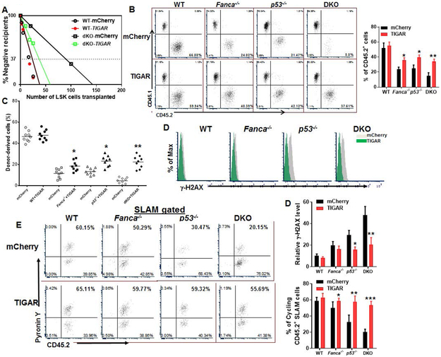
Fig 4. p53-TIGAR ameliorates exhaustion of FA HSCs.
(A) Limited dilution assay. Various doses of sorted transduced LSK cells (25, 75, 150 and 250 mCherry+ cells) expressing mCherry or mCherry-TIGAR were mixed with 2X10⁵ protector BM cells (CD45.1+) and transplanted into irradiated congenic recipients. (n=8-10 per group). Plotted are the percentages of recipient mice containing less than 1% CD45.2+ blood nucleated cells (negative) at 8 weeks after transplantation. Frequency of functional HSCs was calculated according to Poisson statistics. (B) Overexpression of TIGAR improves hematopoiesis reconstitution capacity of FA HSPCs. 1,000 sorted mCherry or mCherry-TIGAR lentiviral vector transduced LSK cells were transplanted into lethally irradiated BoyJ mice. The recipients were subjected to Flow cytometry analysis for donor-derived cells 16 weeks after BM transplantation. Representative flow plots (Left) and quantification (Right) are shown. (n=9 per group). (C) Overexpression of TIGAR improves long-term repopulating activity of FA HSCs. One million BM cells from the primary recipient mice described in (B) were transplanted into sublethally irradiated secondary CD45.1+ recipient mice, and donor-derived chimera in secondary recipients were determined by flow cytometry 16 weeks later. (n=9 per group). (D) Overexpression of TIGAR reduces DNA damage in donor cells. CD45.2+ LSK cells from the recipients described in (B) were subjected to Flow cytometry analysis for γ-H2AX. Representative plots (Upper) and quantification (Lower) are shown. (E) Overexpression of TIGAR increases donor HSC quiescence. Percentage of Pyronin Y+ cells in donor HSC compartment (CD45.2+ SLAM) were analyzed by Flow cytometry. Representative flow plots (Left) and quantification (Right) are shown. (n=6 per group). p<0.05; **, p<0.01;***, p<0.001.

