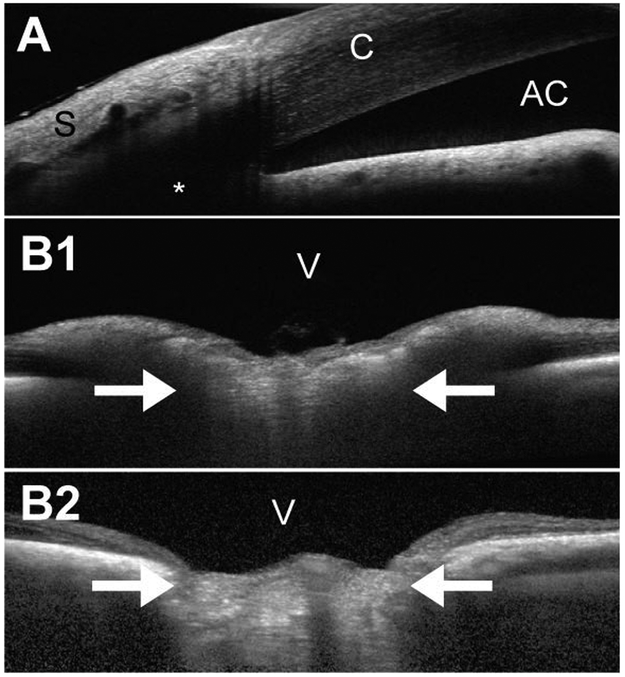
Figure 2.
Optical coherence tomography (OCT) images of the canine eye taken with the Spectralis® (Heidelberg Engineering GmbH, Heidelberg, Germany). (A) Iridocorneal angle of a 2.5-year old, female Beagle with POAG (IOP during imaging: 23 mmHg). OCT often provides higher resolution than routine high-resolution ultrasonography (Fig. 3), but there are still limits when imaging deeper tissues, such as the aqueous humor outflow pathways (*). (B) ONH images of a normal (B1; 6.5-years old female) and POAG-affected (B2; 9.5-years old female) Beagle. While the non-degenerated, well-myelinated normal canine ONH bulges into the vitreous (B1; IOP during imaging: 15 mmHg), the chronically glaucomatous ONH appears cupped (B2; IOP during imaging: 19 mmHg). The white arrows indicate the location of the lamina cribrosa. Unless there is extensive degeneration, the canine lamina cribrosa is difficult to visualize, even with the current enhanced depth imaging (EDI) technology, due to the thick, myelinated prelaminar ONH.
Abbreviations: AC, anterior chamber; C, cornea; S, sclera; V, vitreous.

