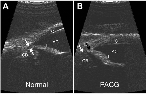
Figure 3.
High-resolution ultrasound (HRUS) images of the canine iridocorneal angle. Compared to a normal eye with physiologic IOP (A) with flat iris and open ciliary cleft (white arrows), the iris has a sigmoidal shape with increased corneal contact (black arrow) and a collapsed ciliary cleft (white arrow) in an eye with acute PACG and IOP of 55 mmHg (B).
Abbreviations: AC, anterior chamber; C, cornea; CB, ciliary body; I, iris.
(From Miller PE 2013 (143); with permission)

