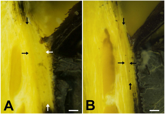
Figure 4.
The canine ciliary cleft may collapse following lens removal by phacoemulsification. Tissue cross-sections of the iridocorneal angle in normal Bouin’s fixed globes show that compared to the normal, unoperated eye (A) the ciliary cleft is severely reduced in an eye 24 hours after phacoemulsification (B). The IOP in this eye reached 52 mmHg 3 hours after surgery and decreased to 15 mmHg at 24 hours. Despite the normalization of IOP, the ciliary cleft remained reduced. The arrows denote the approximate boundaries of the ciliary cleft.
Bars = 0.2mm. (from Miller et al. 1997 (49); with permission).

