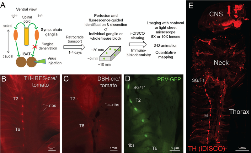
Figure 1.
Overview of experimental design, dissection technique, and whole body tissue clearing. A: Flow diagram of experimental design. B, C: Ventral views of eviscerated, perfused TH:Tomato mouse (B), Dbh:Tomato mouse (C) using stereomicroscope. D: Ventral view of eviscerated, perfused wild-type mouse 96 h post-PRV-GFP injection into the right iBAT pad using stereomicroscope. Note labeling of right, but not left sympathetic chain ganglia. E: Confocal microscope image of the whole upper body of a mouse after iDISCO immunohistochemistry with TH and tissue clearing. Note labeling of the bilateral sympathetic chain, neck nerves, and brain areas. TH, tyrosine hydroxylase; DBH, dopamine beta-hydroxylase; PRV, pseudorabies virus; GFP, green fluorescent protein; SG, stellate ganglion; T1–T7, ganglia for thoracic levels 1–7.

