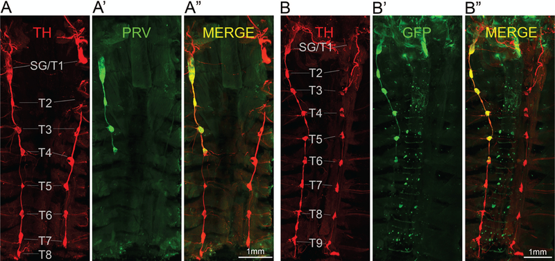
Figure 2.
Ventral overview of sympathetic postganglionic ganglia innervating iBAT in the mouse as shown by two examples with unilateral PRV injections in the right iBAT pad (A, B). Confocal microscope images of tyrosine hydroxylase (TH) (A, B), PRV with GFP expression (PRV-GFP) (A’, B’) and merged images (A”, B”) from iDISCO-processed whole tissue blocks. Note retrograde labeling of the right stellate/T1 ganglion, as well as T2–T4 in example A and T2–T6 in example B.

