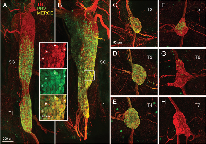
Figure 3.
Postganglionic sympathetic neurons innervating iBAT in the mouse. A, B: Light sheet microscope images of two examples of retrogradely PRV-labeled postganglionic sympathetic neurons in the fused right stellate/T1 ganglion after unilateral PRV injections into the right iBAT pad. Inset shows retrogradely PRV-labeled (green) and unlabeled (red) neurons at higher magnification (asterisks depict examples of co-localized neurons). C-H, Confocal microscope images of individual sympathetic chain ganglia at thoracic levels T2–T7 in the same mouse for which the stellate ganglion is shown in B. Note strong labeling in T2–T4 and much weaker labeling in T5 and T6 ganglia. TH, tyrosine hydroxylase; PRV, pseudorabies virus; SG, stellate ganglion; T1–T7, ganglia for thoracic levels 1–7.

