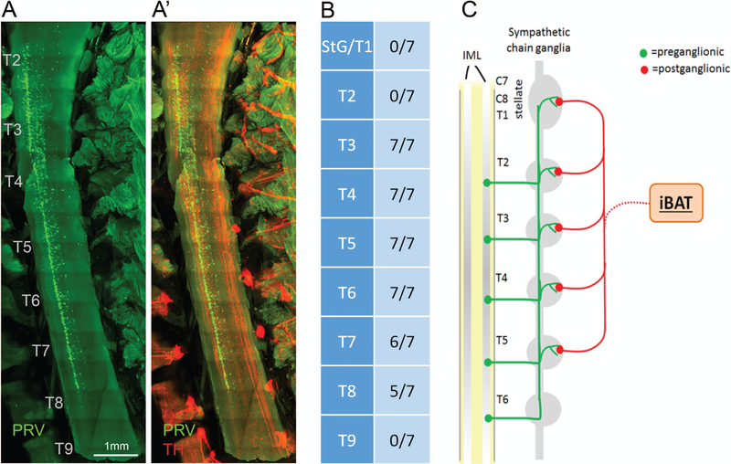
Figure 6.
Location of sympathetic preganglionic neurons innervating iBAT and schematic diagram of sympathetic outflow pattern to iBAT in the mouse. A, A’: Light sheet microscope image through spinal cord showing location of preganglionic neurons (green) on the right side of the intermediolateral column (A), and merged image of PRV and TH (A’) in a mouse with unilateral injection of PRV into the left iBAT pad. B: Semiquantitative assessment of location of postganglionic neurons in the spinal cord relative to the rostrocaudal level. Note the caudal-ward shift in representation with no preganglionic neurons at the level of the fused stellate/T1, T2, and T9 sympathetic chain ganglion. C: Schematic diagram depicting the organization of sympathetic outflow to iBAT in the mouse. IML, intermediolateral column of the spinal cord.

