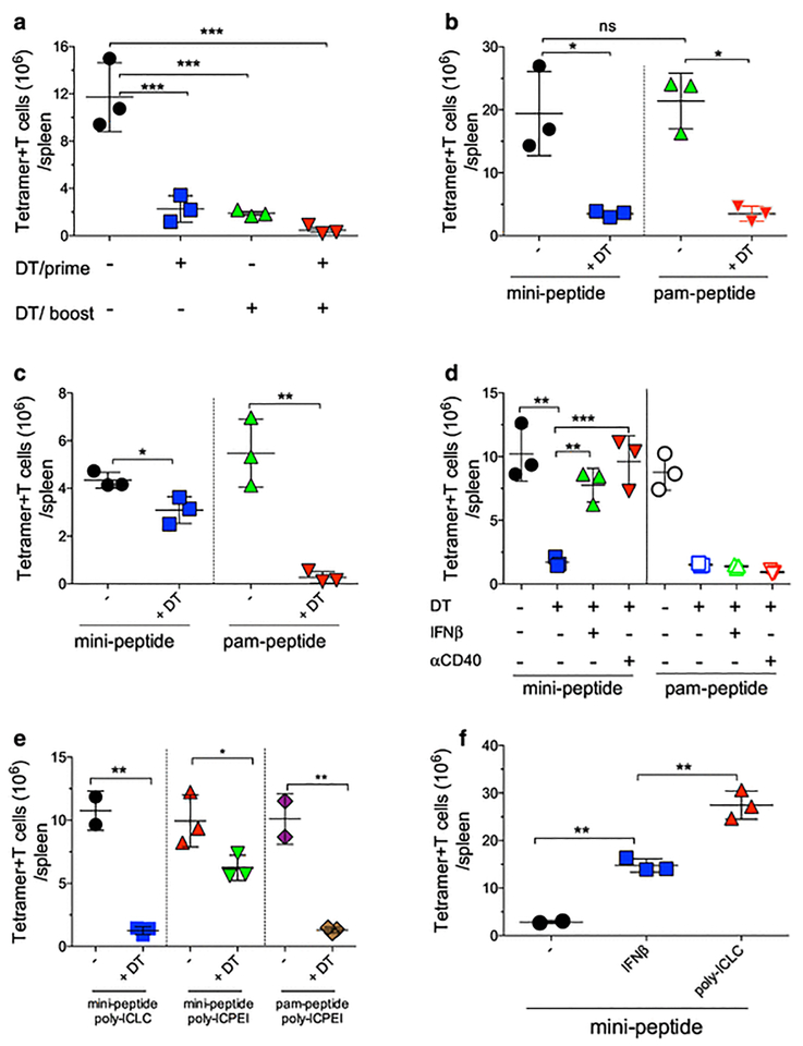
Fig. 1. Role of DCs for T cell expansion induced by peptide vaccination.
a, CD11cDTR BM chimeric mice received 105 naïve OT-I cells followed a day later by prime-boost vaccinations with pam-Ova (pam2-KMVESIINFEKL) and poly-ICLC administered 14 days apart. DCs were depleted with diphtheria toxin (± DT) injections prior to the prime, boost or both as indicated. Numbers of OT-I cells in the spleens were evaluated 7 days after the boost. b, CD11cDTR BM chimeric mice received 105 naïve OT-I cells, were primed with pam-Ova + poly-ICLC and were boosted with pam-Ova or mini-Ova (SIINFEKL) + poly-ICLC. Numbers of OT-I cells in the spleens were determined 7 days after the boost. c Mixed BM chimeric (CD11cDTR and b2M-KO) mice were treated and evaluated as described in b. d, CD11cDTR BM chimeric mice were treated and evaluated as described in b with the addition of twice daily injections of IFNb or one dose of 100 μg aCD40 mAb. e, CD11cDTR BM chimeric mice were treated and evaluated as described in b with boosters using mini-Ova with poly-ICIC or poly-ICPEI or using pam-Ova with poly-ICPEI. f, WT mice received 105 naïve OT-I cells followed by a pam-Ova + poly-ICLC prime and 14 days later were boosted with mini-Ova alone, mini-Ova + 2 daily injections of IFNb or mini-Ova+ poly-ICLC. Results are presented as individual mice (each symbol) with the mean ± SD for each group.

