Fig.2.

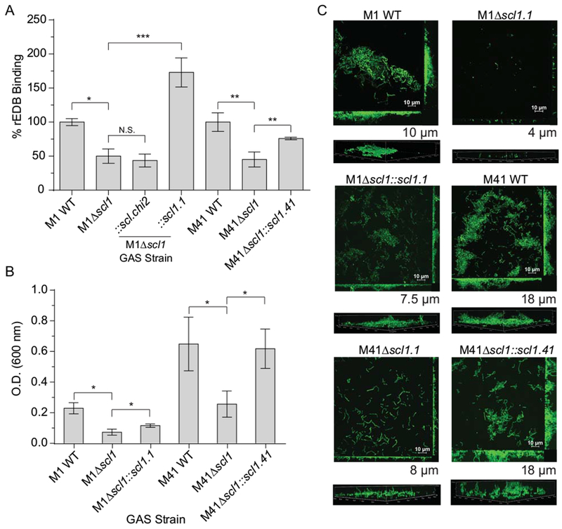
Scl1-EDB binding mediates GAS adherence and biofilm formation. Binding of rEDB to whole GAS cells was compared between WT and Δscl1 mutants, as well as the contribution of surface Scl1 to GAS biofilm formation on rEDB-coated surfaces.
A. rEDB binding to whole GAS cells. Isogenic WT and Δscl1 mutants of the M1- and M41-type GAS strains were used, as well as the M1Δscl1 mutant complemented for the expression of native Scl1.1 (Δscl1::scl1.1) or the chimeric Scl.chi2 (Δscl1::scl.chi2) proteins, and M41Δscl1 mutant complemented for the expression of native Scl1.41 variant (Δscl1::scl1.41). rEDB binding to whole GAS cells was detected by flow cytometry with primary anti-His-tag mAb; binding to GAS WT cells was set as 100%. Statistical analysis was calculated using Student’s two-tailed t-test from three independent experiments (N=3±SD); *P≤0.05, **P≤0.01, ***P≤0.001.
B. Assessment of biofilm formation on rEDB-coated surfaces. M1 and M41 WT, Δscl1 isogenic mutants, and Δscl1 mutants complemented for the expression of native Scl1 variants were compared. Biofilm formation was evaluated spectrophotometrically following crystal violet staining. Graphic bars indicate the mean OD600nm normalized against BSA controls. Statistical analysis was calculated using Student’s two-tailed t-test from three independent experiments (N=3±SD); *P≤0.05.
C. Microscopy imaging of GAS biofilms formed on rEDB coating. The same set of GFP-expressing GAS strains shown in panel B were grown on rEDB-coated glass coverslips for 24 h. Two-dimensional orthogonal views of GAS biofilms are representative of Z stacks from 15 fields over two experiments. Average vertical thickness is indicated in micrometers below two-dimensional orthogonal views, taken from 15 arbitrary fields over two experiments.
