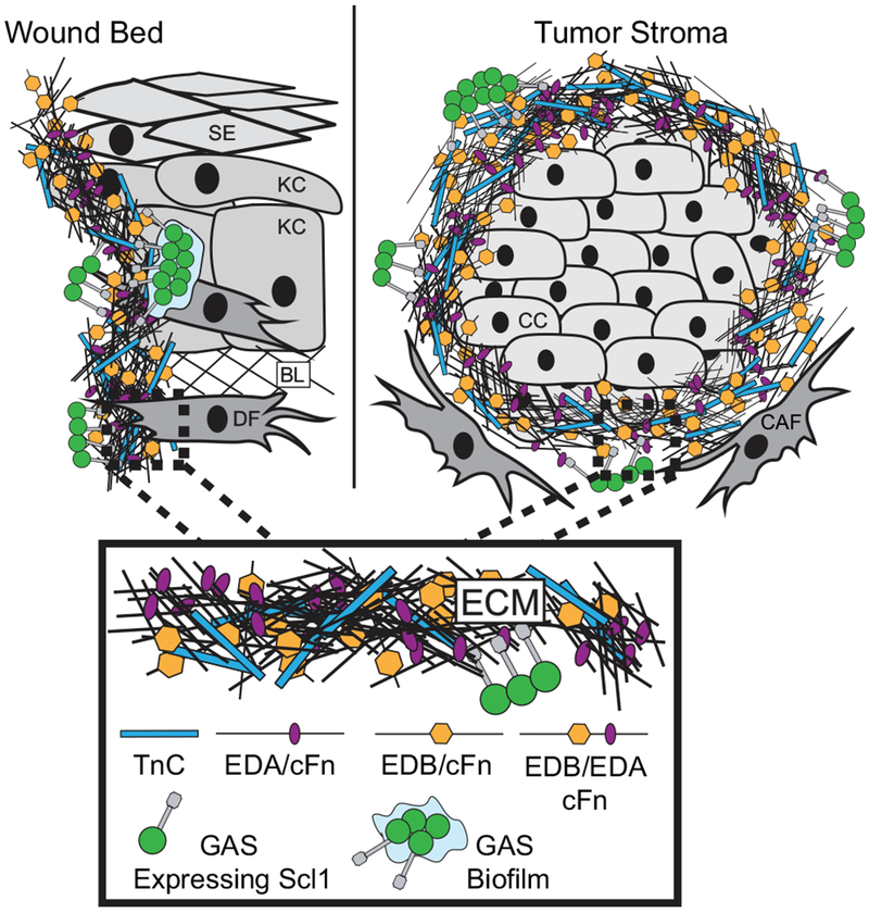
Fig. 8.

Model of GAS colonization of wound and tumor microenvironments. The wound and tumor microenvironments are enriched in isoforms of cellular fibronectin (cFn) that contain extra domain A (EDA) and extra domain B (EDB), as well as tenascin-C (TnC). Left, GAS gains access to the host via portal of entry, such as through a breach in keratinized squamous epithelium (SE), into a tissue environment that contains keratinocytes (KC), basal lamina (BL) ECM, and dermal fibroblasts (DF). Within wound, cells such as DFs deposit cFn isoforms that contain EDA and EDB, as well as TnC. GAS-Scl1 adhesin binds EDA and EDB of cFn, and TnC, promoting call attachment and tissue microcolony formation within the wound. Right, Cancer cells (CC) are surrounded by cancer-associated fibroblasts (CAFs), which deposit cFn isoforms that contain EDA and/or EDB, and TnC, recognized by GAS-Scl1. Enlarged insert, close-up view of the wound- and tumor-associated ECM.
