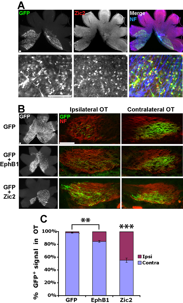
Figure 4. Zic2 induces a greater increase in ipsilateral RGC projections compared to EphB1.
(A) Low power (top) and high power (bottom) images of E18.5 retina electroporated with GFP+Zic2 at E13.5. Note that the majority of GFP+ cells stain positive for Zic2. (B) Representative examples of E18.5 retina and optic tracts from embryos electroporated with GFP, GFP+EphB1 and GFP+Zic2 at E13.5. (C) While electroporating EphB1 at E13.5 did not induce a greater increase in GFP+ ipsilateral projections compared to E14.5 electroporations, ectopic Zic2 expression is significantly more efficient in converting contralateral RGC projections to an ipsilateral fate. n = 5 for GFP control electroporations and n ≥ 10 for EphB1 and Zic2 electroporations, from ≥ 3 different electroporation experiments. Data represent mean ± s.e.m., all scale bars = 100 µm. ANOVA: F(2,25) = 71.28, p < .0001. Modified t-tests: ** = p < .005, *** = p < .0001.

