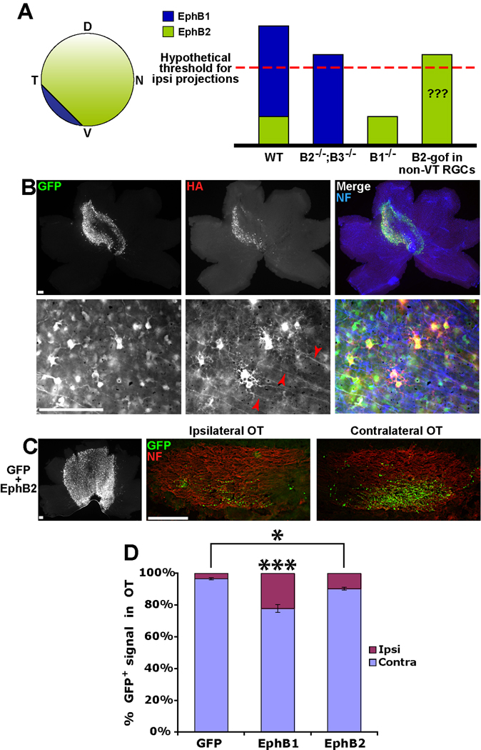
Figure 5. EphB1 induces a greater proportion of GFP+ ipsilateral projections compared to EphB2.
(A) Cartoon depicting expression patterns of EphB1 and EphB2 in the embryonic mouse retina. Graph represents a scenario, consistent with current data from EphB knockout mice, in which the total expression level of EphB receptors exceeds a hypothetical threshold and drives the ipsilateral projection, rather than the specificity of EphB1. We tested whether ectopic expression of EphB2 in RGCs (EphB2 gain-of-function (B2-gof), right column) supports the validity of this model. (B) Low power and high power images of E18.5 retina electroporated with GFP+EphB2 at E14.5. Of note, HA expression is visible on intraretinal RGC axons electroporated with EphB2 (red arrowheads), but HA was rarely observed in EphB1+ axons (Fig. 2A). (C) Representative example of retina and optic tracts electroporated with GFP+EphB2. (D) EphB1 is significantly more efficient at directing ipsilateral projections compared to EphB2 (22% vs. 10%), but EphB2 does induce a greater percentage of uncrossed projections compared to GFP alone (10% vs. 3%). n ≥ 13 embryos for each condition, from ≥ 3 separate electroporation experiments. Data represent mean ± s.e.m., all scale bars = 100 µm. ANOVA: F(2,41) = 31.52, p < .0001. Modified t-tests: * = p < .05, *** = p < .0001.

