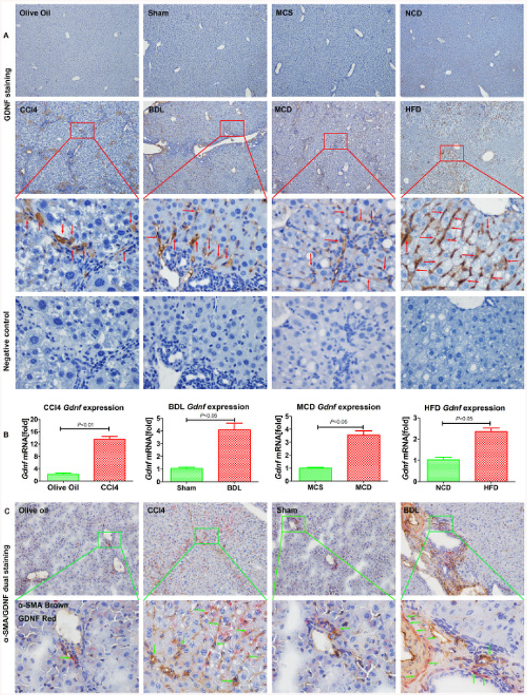
Figure 2.
Glial cell line-derived neurotrophic factor (GDNF) is upregulated in mouse liver fibrosis. (A) Representative images of GDNF staining in mouse liver fibrosis from carbon tetrachloride (CCl4), BDL, methionine-choline-sufficient (MCS), methionine-choline-deficient (MCD), normal chow diet (NCD), high fat diet (HFD) samples. Original magnification x100, the third line is x600, fourth line is the negative control showing a serial section of the third line x600, the red arrow indicates GDNF-positive staining, n=5 per group. (B) Gdnf mRNA expression in whole liver of CCl4, BDL, MCD and HFD mice. Bars indicate the mean±SD of three independent experiments; n=5 per group; the t-test with the non-parametric Mann-Whiney U test was used. (C) Frozen mouse liver sections on dual α-SMA and GDNF immunohistochemistry (original magnification x100), the lower part represents the boxed area with x600 magnification, the green arrow indicates α-SMA and GDNF dual positive staining, n=5 per group.

