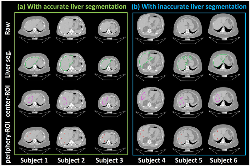
Figure 4.
Qualitative visualizations of raw abdomen CT scan, liver segmentation, center-ROI and periphery-ROI from ALARM pipeline (a) shows the results of three subjects from accurate liver segmentation, while (b) presents the results of three subjects with the inaccurate liver segmentation. The first row indicates the raw input CT scan. The second row shows the liver segmentation from the deep learning segmentation. The third row shows the central-ROI, while the fourth row shows the periphery-ROI. The ROI based liver attenuation method is able to tolerate imperfect whole liver segmentation after performing morphological operations

