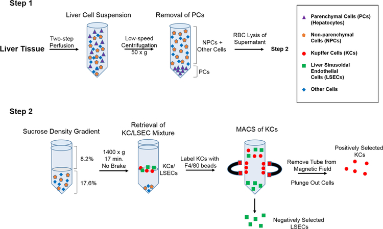
Figure 1. Isolation and enrichment protocol for KCs and LSECs from mouse livers.
In Step 1, liver tissues were subjected to a retrograde two-step perfusion followed by low speed centrifugation to remove hepatocytes. The RBCs were then lysed with ammonium chloride and the remaining cell mixture was subjected to sucrose density gradient centrifugation. In Step 2, the KC/LSECs populations partition was collected and mixed with anti-F4/80 antibody-coated magnetic beads. KCs were positively selected by magnetic separation, while LSECs were enriched in the flow through. Relative enrichment and viability of primary KCs and LSECs was determined by flow cytometry and phase contrast microscopy.

