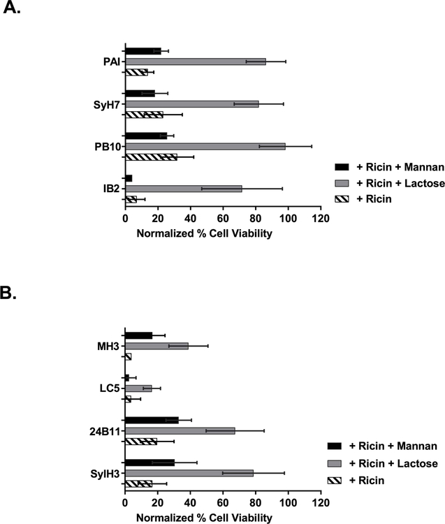
Figure 9. Anti-ricin MAbs function via the mannan-dependent uptake pathway in LSECs.
Freshly isolated LSECs were seeded in 96 well microtiter plates and pulsed for 2 h with ricin plus indicated anti-RTA (panel A) or anti-RTB (panel B) MAbs, in the presence of α-mannan (1 mg/mL) or lactose (0.1 M). Cell viability was normalized to LSECs that had not been treated with ricin toxin. In all cases, the addition of lactose resulted in a significant increase in cell viability, as compared to as compared to control (ricin only). Mannan addition impacted cell viability in only one instance (MH3). Kruskal-Wallis with Tukey’s multiple comparisons test (GraphPad 7) was applied to account for non-normal distribution of data.

