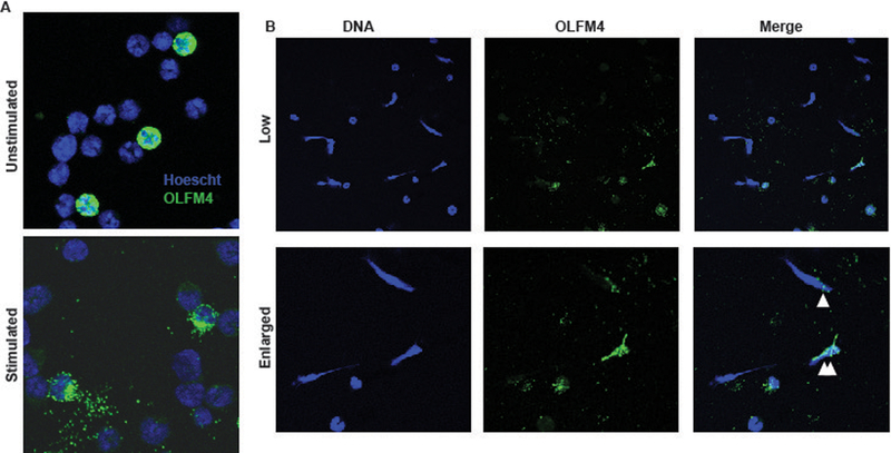
Figure 4. Murine OLFM4 is not associated with neutrophil NETs.

A. Immunofluorescence of purified bone marrow neutrophils from BalbC mice using Hoescht dye to stain DNA (blue) and Af488 to stain OLFM4 (green) showing approximately 20% OLFM4+. Following stimulation with PMA, OLFM4 staining can be seen more toward the periphery of the activated neutrophil. B. Immunofluorescence staining of low magnification (60x; top) and enlarged (bottom) magnification of stimulated neutrophils showing formation of NETs. Single white triangle designates and OLFM4 negative neutrophil that has a few small areas of OLFM4 colocalized staining. Double white triangles designate and OLFM4+ neutrophil that has undergone NETosis.
