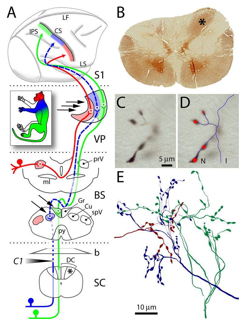
Fig. 1.
Dorsal column lesion and tract-tracing. (A) The lemniscal and trigeminal pathways convey topographically organized somatosensory information (color-coded as in inset) from large-diameter primary sensory axons on the left side of the body to the right thalamus (VP) and cortex (S1). Lesion of the left cuneate fasciculus at the C1 level of the spinal cord (SC) (shaded arrowhead) caused Wallerian degeneration of primary sensory axons above the lesion (dotted line). Dashed lines show the component of the pathway deafferented by the lesion. Arrows point to dorsal column nuclei and VP sites where BDA was injected. b, plane of cut of the section shown in B; asterisks denote normal cuneate fasciculus. (B) Transverse section from SC immunostained for parvalbumin, showing loss of axons in the left cuneate fasciculus, compared to the right (asterisk). (C) Anterogradely labeled axon terminals in VP at high magnification. Only the portion in sharp focus is traced at any given time. (D) Line rendering of Neurolucida tracing of the same axon through the thickness of the section. Red circles mark synaptic boutons (varicosities). Line and marker size were continuously updated to match the size of axons and varicosities. (E) Cluster of three axons terminating in VP, reconstructed from multiple consecutive sections. Abbreviations: CS, central sulcus; Cu, cuneate nucleus; DC, dorsal columns; Gr, gracile nucleus; I, incomplete ending; IPS, intraparietal sulcus; LF, longitudinal fissure; LS, lateral sulcus; ml, medial lemniscus; N, normal ending; prV, principal trigeminal nucleus; py, pyramidal decussation; spV, spinal trigeminal nucleus; VP, ventral posterior nucleus.

