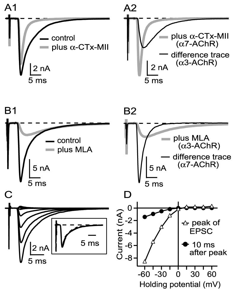
Figure 1.
Evoked EPSCs elicited from ciliary neurons have contributions from both α3-nAChRs and α7-nAChRs (A-B). α3-nAChR and α7-nAChR mediated currents show inward rectification (C-D). A1 and B1 show examples of EPSCs elicited by 0.03 Hz preganglionic stimulation before and after addition of either 300 nM α-CTx MII (A1), which blocks α3-nAChRs, or 50 nM MLA (B1), which blocks α7-nAChRs. A2 reproduces the EPSC from A1 in α-CTx MII (α7-nAChR current) plus a difference curve (before minus after, representing α3-nAChR current), while B2 reproduces the EPSC in B1 in MLA (α3-nAChR current) plus a difference curve (representing α7-nAChR current). α3-nAChR currents are slower than α7-nAChR currents (A2, B2). All records are the average of 3-5 traces. C shows an EPSC from one connection at holding potentials of -60 mV to +60 mV (15 mV increments), and D shows the current at the peak (dominated by α7-nAChRs) and 10 ms after the peak (dominated by α3-nAChRs) vs. holding potential, uncorrected for a calculated -5 mV liquid junction potential . Both α7-nAChR- and α3-nAChR-mediated currents rectify to about the same degree. Each trace in C is the average of 3 traces collected at 0.03 Hz. Similar results were obtained in three other cells.

