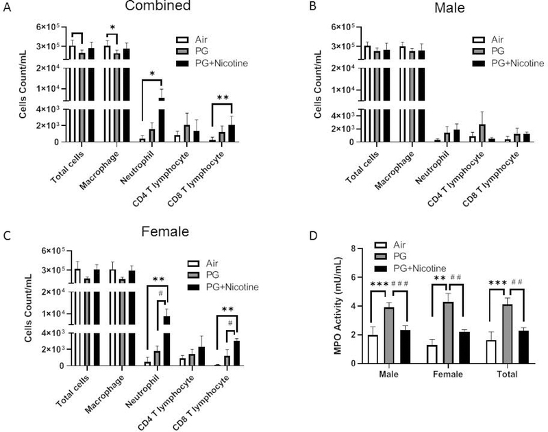
Fig. 1. Acute exposure to inhaled e-cig aerosols containing nicotine increases inflammatory cellular influx in BALF.
Mice were exposed to air, PG, and PG with nicotine (PG+Nicotine) for 3 days (2 hrs/day). Mice were sacrificed 2 hrs post-final exposure on day 3. Total cell counts per milliliter in BALF were determined by trypan blue staining using Bio-Rad Tc10 cell counter. Differential cells: F4/80+ macrophages, LY6B.2+ neutrophils, CD4a+ T-lymphocytes, and CD8a+ T-lymphocytes were determined by flow cytometry, combined results (A), male (B) and female (C) were presented individually. (D). MPO activity in BALF. Data are shown as mean ± SEM (n=6/group; equal number of male and female mice were used; n=3 for male and female groups respectively; *P < 0.05, **P < 0.01, significant compared with air exposed control; # #P < 0.01, # # #P < 0.001 significant compared with PG exposed group.)

